Subdural Hematoma: Understanding Its Causes, Treatments, and Prognosis
Subdural hematomas (SDHs) are a type of brain injury that occurs when blood accumulates in the space between the brain and the skull, typically due to the rupture of bridging veins. This article provides an overview of the symptoms, causes, diagnosis, and treatments of acute, subacute, and chronic SDHs.
---
### Symptoms
SDHs can be categorised into three types based on symptom onset: acute, subacute, and chronic.
Acute SDHs develop rapidly, with symptoms such as altered mental status (confusion, decreased consciousness), focal neurological signs (weakness on one side, slurred speech), signs of increased intracranial pressure (headache, nausea, vomiting), seizures, and possible progression to coma and brain herniation if left untreated.
Chronic SDHs, on the other hand, develop gradually, often over weeks, with symptoms like cognitive deficits (memory impairment, confusion), personality changes, and gradual onset of focal neurological impairments. These symptoms can be mistaken for dementia or stroke, especially in elderly patients or those on blood thinners.
Subacute SDHs exhibit features overlapping with both acute and chronic forms, with symptom onset between the acute and chronic phases.
---
### Causes
The most common cause of SDHs is head trauma, ranging from minor bumps or falls to severe injuries such as assaults or accidents. Other risk factors include cerebral atrophy (common in elderly individuals), coagulopathies, hypertension, and the use of anticoagulants or antiplatelet drugs. The rupture of bridging veins between the dura and arachnoid mater leads to bleeding into the subdural space.
---
### Diagnosis
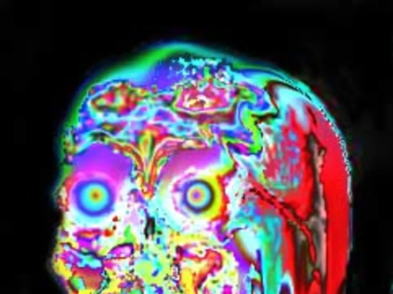
Noncontrast head CT is the diagnostic modality of choice for SDHs. It typically shows a crescent-shaped hematoma that can cross cranial sutures. Acute SDH appears hyperdense, while chronic SDH appears hypodense or isodense depending on age.
---
### Treatment
Treatment options for SDHs depend on the type and severity of the hematoma.
Acute SDHs often require emergency neurosurgery (craniotomy or burr hole evacuation) if the hematoma is ≥10 mm or if there is a midline shift ≥5 mm, or if the patient is symptomatic. Rapid intervention is critical due to high mortality rates (up to ~20-50%, higher if combined brain injury).
Subacute and chronic SDHs may be treated with surgical drainage (burr hole drainage) for symptomatic cases, or conservative management for small, asymptomatic SDHs with no ICP signs.
Emerging medical management options, such as atorvastatin plus low-dose dexamethasone, show promise in some acute SDH cases but are not yet standard care.
---
### Summary
In summary, SDHs are a serious medical condition that can lead to death or permanent brain damage if left untreated. Acute SDHs are medical emergencies, while chronic SDHs develop gradually and may be mistaken for other neurological conditions. Treatment depends on the type and severity of the hematoma, with emergency surgery often required for acute cases. Emerging medical management options offer hope for patients who refuse surgery or have contraindications to surgical intervention.
- Other neurological disorders may exhibit similar symptoms as subdural hematomas, making prompt recognition and effective treatment crucial for health-and-wellness.
- The causation of subdural hematomas is primarily associated with head trauma, but coagulopathies, hypertension, and the use of anticoagulants or antiplatelet drugs can also contribute to these neurological-disorders.
- In managing subdural hematomas, the medical-conditions, including the type and severity of the hematoma, play a significant role in determining if emergency surgery or conservative methods like blood drainage are necessary for a patient's fitness-and-exercise.
- Neuroscience and the practice of medicine continue to evolve, with ongoing research into new treatments for subdural hematomas, such as the use of atorvastatin and dexamethasone, that may provide alternatives to invasive surgeries for some patients.
- Blood tests and scans such as CT scans are crucial in diagnosing subdural hematomas, aiding healthcare providers in making informed decisions on appropriate treatments for these neurological medical-conditions.


